This Website uses Cookies

Cookies are managed according to our Privacy Policy

Neural Correlates of the Consciousness of Fear

Tiny Blue Dot Foundation | Massachusetts Gneral Hospital logo

Overview:

This project aimed to understand the brain mechanisms responsible for fear and how conciousness plays a role in the fear factor.

Abstract:

Fear is one of the most fundamental motivating factors in human life. The neurobiology of fear has been extensively studied both in animals and humans. What is largely unknown, however, is how specific nodes of the fear circuitry contribute to the conscious experience of fear. 

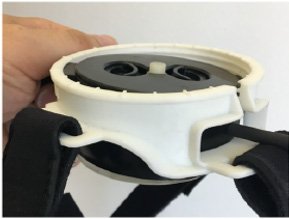
This project involves probing the brain regions involved in the conscious experience of fear. Probing will be accomplished using a novel, noninvasive form of brain stimulation, transcranial Focused Ultrasound (tFUS)/Low Intensity Focused Ultrasound Pulsation (LIFUP), in conjunction with functional Magnetic Resonance Imaging (fMRI). 

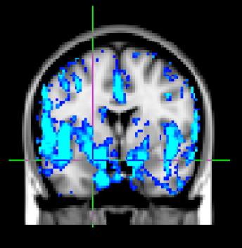
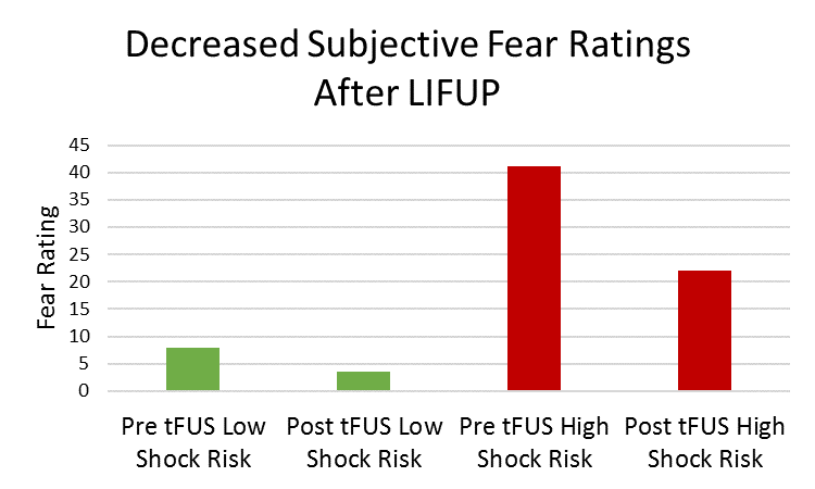
We hypothesize that, during a fear-inducing task, active tFUS of the amygdala will decrease activity within the fear circuit and correspond with lowered skin conductance ratings and fear ratings, compared with sham tFUS. We have enrolled n=16 healthy controls in this study thus far. 

Tiny Blue Dot Foundation | Neural Correlates Fig 1a GraphicTiny Blue Dot Foundation | Neural Correlates Fig 2aGraphic
Tiny Blue Dot Foundation | Neural Correlates Fig 2b Graphic
Tiny Blue Dot Foundation | Neural Correlates Fig 3a Graphic
Tiny Blue Dot Foundation | TNeural Correlates Fig 3b Graphic


Broader Impact:

The results from this study have larger implications for the conscious experience of fear and tFUS/LIFUP. Specifically, tFUS/LIFUP of the amygdala will elucidate the causal role of the amygdala in objective and subjective components of fear. If tFUS/LIFUP can decrease subjective fear, this suggests that tFUS of the amygdala has therapeutic potential for individuals with clinical levels of fear/anxiety, such as anxiety disorders and specific phobias.

Publications [08]

  • Y. Tu, et al., Manipulating placebo analgesia and nocebo hyperalgesia by changing brain excitability. Proc Natl Acad Sci USA 118, e2101273118 (2021).

  • A.S. Widge, et al., Deep brain stimulation of the internal capsule enhances human cognitive control and prefrontal cortex function. Nat Commun. 10, 1536 (2019).

  • K.K. Ellard, et al., Intrinsic functional neurocircuitry associated with treatment response to transdiagnostic CBT in bipolar disorder with anxiety. Journal of Affective Disorders 238, 383-391 (2018).

  • D.D. Dougherty, T. Chou, U. Buhlman, S.L. Rauch, T. Deckersbach, Early amygdala activation and later ventromedial prefrontal cortex activation during anger induction and imagery. Journal of Medical Psychology 22, 3-10 (2017).

  • R.T. Faghih, et al., Characterization of fear condition and fear extinction by analysis of electrodermal activity. Annu Int Conf IEEE Eng Med Biol Soc.7814-8 (2015).

  • M.F. Marin, J.A. Camprodon, D.D. Dougherty, M.R. Milad, Device-based brain stimulation to augment fear extinction: implications for PTSD treatment and beyond. Depress Anxiety, 31, 269-78 (2014).

  • H. Wey, et al., Simultaneous fMRI-PET of the opiodergic pain system in human brain. Neuroimage 102, 275-82 (2014).

  • M.A. Zeidan. Test-retest reliability during fear acquisition and fear extinction in humans. CNS Neurosci Ther. 18, 313-7 (2012).